Dr. Yongsheng Chen’s Laboratory
Quantitative MRI (qMRI) of the Brain and Peripheral Nerves
Dr. Chen’s research is focused on developing quantitative MRI (qMRI) methods of the brain and peripheral nerves. His clinical research interests include neurodegenerative disorders (e.g. Parkinson’s disease and Multiple Sclerosis) and peripheral neuropathies. In collaboration with his colleagues, Dr. Chen has developed a rapid multiparametric qMRI method of the brain referred to as strategically acquired gradient echo (STAGE) imaging. STAGE shortens the time span in studying each patient and still acquires the desired quantitative and qualitative images of the brain. One of his recent contributions to science is the development of a multiparametric qMRI method for peripheral nerves in the leg. The nerve qMRI method has been used to systematically assess the demyelination and axonal loss of peripheral nerves in patients with polyneuropathies. His research laboratory is currently funded by the NIH and Wayne State to develop monitoring biomarkers in patients with inherited peripheral nerve diseases, and diagnostic biomarker signatures to differentiate demyelination from axonal degeneration types of polyneuropathies.
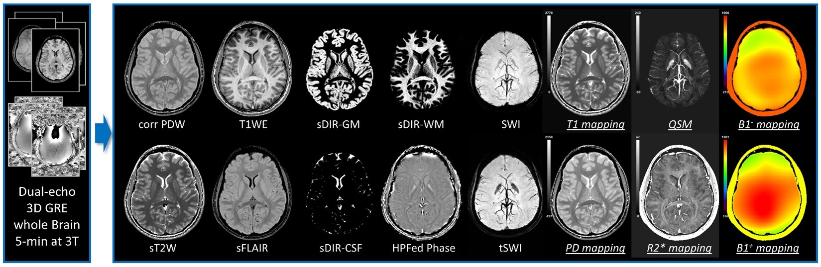
STAGE Imaging employes multi-echo multi-flip-angle gradient echo sequences to acquire whole-brain covered qualitative and quantitative data in a rapid manner [Chen Y et al., MRI 2018; Wang Y et al., MRI 2018; Haacke EM et al., MRI 2020]. The representative data in this figure was acquired in 5 minutes at 3T covering the whole brain. The results include qualitative images such as PDW, T1W, simulated T2W (sT2W), s FLAIR, sDIR images for GM, WM and CSF, as well as quantitative data such as T1, T2*, proton density mappings and quantitative susceptibility mappings.
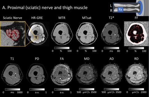
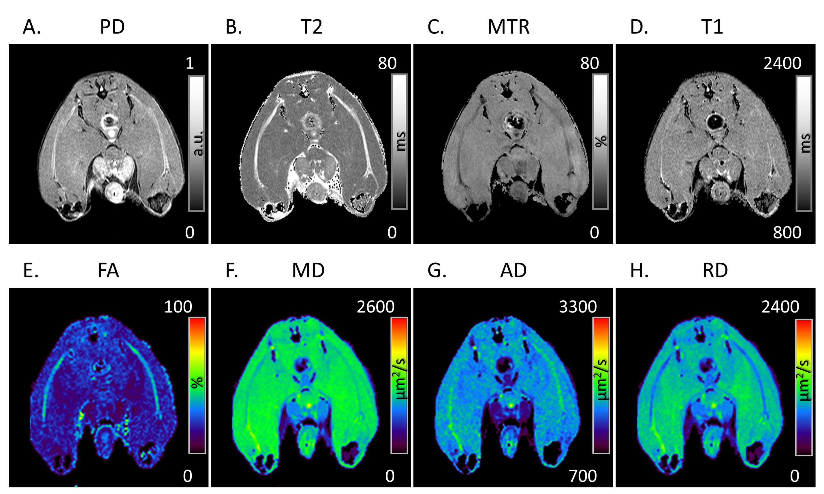
Multiparametric quantitative MRI to systematically assess the pathologies of the muscles and peripheral nerves in the leg in patients with polyneuropathies [Chen Y et al, F1000Res 2019; Chen Y et al., JMRI 2021; Fritz N et al., ACTN 2020; Saba S et al., ACTN 2020; Chen Y et al., JMRI 2023]. The qMRI protocol quantifies the morphologies of the sciatic nerve and tibial nerve with ultra-high resolution 3D MRI, and quantitative maps of MTR, MT saturation index, T2*, T1, proton density, FA, and mean/axial/radial diffusivities. In addition, the method evaluates the denervation and intramuscular fat infiltration of skeletal muscles using the above quantitative maps plus the fat fraction map of the muscle.
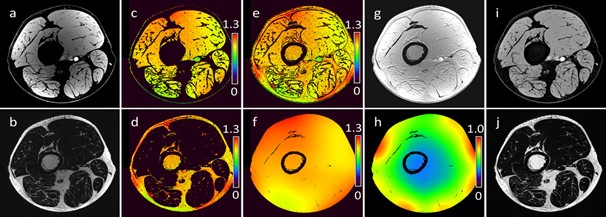
Corrections of RF field inhomogeneities using fitting based method [Chen Y et al., JMRI 2021]. The method estimates the RF transmission field variation (B1+) and the receiver coil sensitivity map (B1-) and corrects them separately for quantifying the T1, proton density, and magnetization transfer ratio.
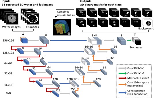
Automation of quantifying axonal loss in patients with peripheral neuropathies through deep learning derived muscle fat fraction [Chen Y et al., JMRI 2021]. We trained a deep learning-based model for automated segmentation and quantification of individual muscles using a 3D U-Net architecture, to facilitate the efficiency and accuracy of analyzing patient data in clinical studies to develop monitoring biomarkers in patients with peripheral neuropathies.
Multiparametric qMRI of sciatic nerves in a Pmp22 heterozygous knockout mouse model, which is for mimicking a human disease called HNPP, to determine the molecular substrates responsible for these qMRI readouts [Chen Y et al., PNS Annual Meeting 2022; Chen Y et al., ANA Annual Meeting 2022].
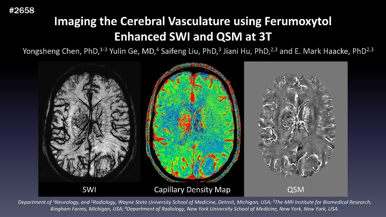
Imaging the Cerebral Vasculature using Ferumoxytol Enhanced SWI and QSM at 3T [Chen Y et al., ISMRM 2019]. The iron-based MRI contrast agent in line with the high-resolution SWI imaging enhances the visualization of the cerebral vasculature in a micro-level. The method also makes it possible to visualize micro-level cerebral arteries at 3T in humans in vivo.